
The distinction between a congenital and hereditary defect is not about assigning blame; it is the critical first step in defining your pet’s prognosis and management plan.
- Congenital simply means “present at birth” and can be caused by environmental factors, while hereditary means it is genetically transmissible. A defect can be both.
- Effective management, from surgery to lifestyle changes, depends on an accurate diagnosis of the specific condition, not its origin.
Recommendation: Use this clinical framework to partner with your veterinarian, ask the right questions, and build a proactive care strategy for your pet’s future.
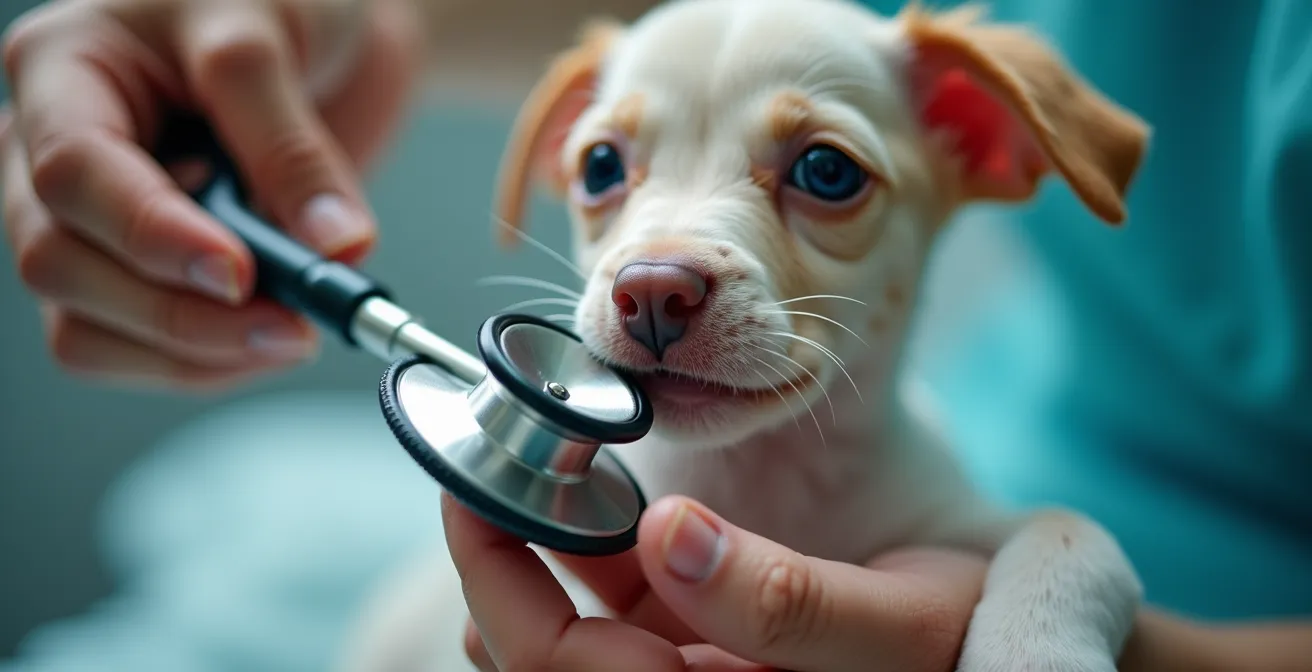
The moment a veterinarian places a stethoscope on your new puppy’s chest and their expression shifts is one of profound silence. That quiet “whoosh” of a heart murmur, or the subtle limp that doesn’t resolve, triggers a cascade of questions for an owner. The most pressing of these often circles around guilt and confusion: “Is this my fault? Was this bad luck, or was it bad breeding?” This leads to the clinical terms ‘congenital’ and ‘hereditary,’ which are frequently used interchangeably but have distinct and critical implications.
From a pathological standpoint, understanding this difference is paramount. A congenital disorder is one that is present at birth. It may be inherited, but it could also be the result of an environmental influence on the fetus during development. A hereditary disorder, however, is specifically caused by a genetic mutation passed down from the parents. Therefore, all hereditary disorders are congenital, but not all congenital disorders are hereditary. This is more than semantics; it is the foundation of a sound diagnostic and prognostic pathway.
This article moves beyond the blame game. It provides a clinical framework for understanding your pet’s condition. We will not just define terms but explore the real-world management decisions they influence—from surgical timing and insurance coverage to the ethical imperative of spaying. By dissecting specific conditions through this lens, you will be empowered to navigate the path forward with clarity and confidence, transforming your role from a worried owner to a proactive partner in your pet’s lifelong care.
To help you navigate these complex topics, this guide is structured to address the most critical questions and decisions you will face. We will examine specific defects and the management strategies they require, providing a clear roadmap for your journey.
Table of Contents: A Clinical Guide to Your Pet’s Defect
- BOAS Surgery: When to Operate to Improve Breathing?
- Conservative Management for Dysplastic Puppies: Can You Avoid Surgery?
- Living With a PDA: The Prognosis With and Without Repair
- Pre-Existing Conditions: Will Insurance Cover a Congenital Flaw?
- Spaying a Dog With a Defect: Why It Is Non-Negotiable?
- OFA vs. PennHIP: Which Predicts Arthritis Risk More Accurately?
- Surgery or Conservative Management: Deciding for a Torn CCL
- Glucosamine vs. Green Lipped Mussel: Which Actually Repairs Joints?
BOAS Surgery: When to Operate to Improve Breathing?
Brachycephalic Obstructive Airway Syndrome (BOAS) is a classic example of a hereditary condition. The traits we have selected for in flat-faced breeds—a shortened skull and compressed nasal passages—are directly responsible for this debilitating breathing disorder. The question is not if the condition is genetic, but when the clinical signs have progressed to a point that surgical intervention becomes necessary for quality of life. The decision to operate is a balance of risk and reward.
Timing is critical. Operating too early on a dog with mild signs may be unnecessary, but waiting too long can lead to secondary, irreversible changes like laryngeal collapse. Key indicators for surgery include significant exercise intolerance, frequent gagging or regurgitation, and episodes of sleep apnea. Documenting these events provides your veterinarian with objective data to guide the decision. It is a clinical reality that these dogs face higher anesthetic risks; in fact, research from 2024 shows brachycephalic dogs are 4.33x more likely to have post-anesthetic complications. However, this risk can be mitigated.
A multi-level surgical approach—addressing stenotic nares (narrow nostrils), an elongated soft palate, and everted laryngeal saccules—can dramatically improve airflow. A 2024 retrospective study of 199 dogs confirmed the benefits but also highlighted risks, with aspiration pneumonia being a primary concern, especially in overweight patients. Therefore, pre-surgical conditioning is not just recommended; it is a critical part of the management pathway. Implementing a weight management program and addressing underlying gastrointestinal issues, which affect up to 80% of BOAS dogs, significantly improves the chances of a successful outcome.
Conservative Management for Dysplastic Puppies: Can You Avoid Surgery?
Hip dysplasia is a malformation of the hip’s ball-and-socket joint, a condition with strong hereditary links but also influenced by environmental factors like rapid growth and nutrition. For a dysplastic puppy, the clinical pathway diverges into two main routes: surgical correction or conservative management. The idea of avoiding surgery is appealing, but it is not a suitable option for every case. The decision hinges on the puppy’s age, the severity of the joint laxity, and the presence of osteoarthritis.
Early diagnosis is key. Procedures like Juvenile Pubic Symphysiodesis (JPS) must be performed at 12-16 weeks of age, while a Triple Pelvic Osteotomy (TPO) is best for dogs under 10 months without any arthritic changes. For mild cases or for dogs that are not surgical candidates, conservative management becomes the primary strategy. This is not a passive approach; it is an active, multi-faceted plan aimed at preserving joint function and managing pain. The core components include weight control, physical therapy, and strategic home modifications to reduce stress on the joints.
Creating a joint-sparing environment is a non-negotiable part of conservative care. This involves practical changes that minimize the daily impact on your puppy’s developing hips. Implementing these modifications can significantly improve comfort and may delay the progression of arthritis, extending the window before more aggressive interventions are needed.
Action Plan: Creating a Joint-Sparing Environment
- Install non-slip rugs or mats on all hard flooring surfaces to prevent slipping.
- Build or purchase ramps to help your dog access furniture and vehicles safely.
- Elevate food and water bowls to reduce strain on the neck and front-end joints.
- Replace high-impact activities like jumping for a ball with controlled leash walks or swimming.
- Use baby gates to prevent unsupervised access to stairs, which puts significant force on the hips.
Living With a PDA: The Prognosis With and Without Repair
The discovery of a heart murmur in a puppy often leads to a diagnosis of Patent Ductus Arteriosus (PDA). This is a classic congenital defect. The ductus arteriosus is a blood vessel that is essential for fetal circulation in the womb, allowing blood to bypass the lungs. It is supposed to close within hours or days after birth. When it fails to close, it is “patent,” creating a left-to-right shunt of blood that overloads the heart and lungs. While it has a known hereditary basis in certain breeds like Poodles and Maltese, it can also occur spontaneously.
The prognosis for a puppy with a PDA is starkly different depending on the chosen clinical pathway. Without intervention, the constant volume overload on the heart leads to congestive heart failure. Most untreated dogs will not survive past their first year. The decision is therefore less about “if” to intervene and more about “how” and “when.” The evidence is definitive: surgical repair dramatically improves outcomes with a 92% survival rate, compared to only 36% for dogs managed with medication alone.
The repair effectively cures the condition. This can be achieved through traditional open-chest surgery to ligate (tie off) the vessel or, increasingly, through minimally invasive procedures where a device is placed via catheter to occlude the PDA. The choice of technique depends on the patient’s size and the veterinarian’s expertise. The key takeaway is that a PDA diagnosis, while frightening, is one of the most correctable congenital heart defects. A positive outcome is highly likely with timely surgical correction, allowing the puppy to live a completely normal life.
Pre-Existing Conditions: Will Insurance Cover a Congenital Flaw?
Navigating pet insurance is a complex task, made even more so by the presence of a congenital or hereditary condition. The fundamental rule of nearly all insurance policies is that they do not cover pre-existing conditions. If your pet shows clinical signs of a defect *before* the policy’s effective date and waiting period have passed, any related diagnostics or treatments will almost certainly be excluded from coverage. This is where the line between a breeder’s responsibility and an owner’s financial burden becomes sharply defined.
However, the situation is different for conditions that are diagnosed *after* the policy is in effect. Many modern insurance providers now offer coverage for hereditary and congenital conditions, provided they were not apparent before enrollment. This is a crucial distinction. For example, if you insure a puppy that seems perfectly healthy, and it is later diagnosed with hip dysplasia at one year of age, the treatment may be covered. This highlights the importance of securing insurance as early as possible in your pet’s life, before any underlying issues have a chance to manifest.
To protect yourself and ensure potential coverage, a proactive documentation strategy is essential. A comprehensive veterinary examination immediately upon acquiring your pet establishes a clean bill of health at policy inception. For breed-specific issues, performing screening tests like preliminary hip radiographs or a cardiac evaluation *before* a policy starts can document the absence of disease. Keeping meticulous records, including the health clearances of the puppy’s parents, creates a paper trail that can be invaluable in the event of a future claim. This is not about trying to “beat” the system; it is about establishing a transparent and well-documented health history from day one.
Spaying a Dog With a Defect: Why It Is Non-Negotiable?
The decision to spay or neuter a pet with a diagnosed hereditary defect is not a lifestyle choice; it is an ethical imperative. While congenital defects occur in an estimated 0.2% to 3.5% of all canine births, those with a known genetic basis carry a responsibility to halt their propagation. Allowing an animal with a deleterious genotype to reproduce is the primary mechanism by which these painful and costly diseases become more prevalent within a breed. It knowingly passes the problem to a new generation of dogs and their owners.
From a pathologist’s perspective, hereditary diseases are a matter of population genetics. As explained by Dr. Jerold Bell’s research at the AVMA Virtual Convention 2020, conditions like hip dysplasia, many inherited cancers, and mitral valve disease persist due to the accumulation of disease liability genes in the breeding pool. When breeders do not actively select against the phenotype (the observable traits of the disease), the underlying genes are passed on. Responsible breeding is defined by rigorous health screening and the removal of affected animals—and their close relatives—from breeding programs.
For the owner of a pet with a single defect, the decision might seem isolated. However, it is part of a larger ecosystem of breed health. Spaying or neutering is the single most effective action an individual owner can take to be part of the solution. It ensures that a known genetic flaw, whether it’s for a severe condition like degenerative myelopathy or a “manageable” one like certain allergies, stops with their beloved pet. It is a definitive, responsible act that prioritizes the future health and welfare of the breed over any other consideration.
OFA vs. PennHIP: Which Predicts Arthritis Risk More Accurately?
When hip dysplasia is suspected, objective diagnosis is the next step. Two primary methods are used for screening: the Orthopedic Foundation for Animals (OFA) and the University of Pennsylvania Hip Improvement Program (PennHIP). While both aim to assess hip quality, they are fundamentally different tools designed to answer different questions. Understanding their methodologies is key to interpreting their results and predicting future risk. The choice between them depends on the owner’s goal: breeding certification or early risk assessment for a pet.
The OFA method is a static assessment. It requires the dog to be at least 24 months old and involves a single X-ray taken in a specific “hip-extended” position. A panel of radiologists then subjectively grades the hips as Excellent, Good, Fair, Borderline, or various degrees of dysplastic. Its primary utility has historically been as a pass/fail system for breeding certification.
PennHIP, in contrast, is a dynamic stress test that can be performed as early as 16 weeks of age. It measures passive hip laxity—the root cause of dysplasia—by taking radiographs in three different positions. The result is a quantitative Distraction Index (DI) score, which is an objective measure of how loose the hips are. This score directly correlates to the likelihood of developing osteoarthritis later in life. A DI score closer to 0 indicates very tight hips, while a score closer to 1.0 indicates extreme laxity. This method is not for certification, but for prognostic risk stratification. The table below outlines the key differences.
| Aspect | OFA | PennHIP |
|---|---|---|
| Minimum Age | 24 months (2 years) | 16 weeks |
| Assessment Type | Static ‘best pose’ snapshot | Dynamic passive laxity stress test |
| Results Format | Subjective grades (Excellent/Good/Fair) | Quantitative Distraction Index (DI) score |
| Primary Use | Breeding certification | Early intervention planning |
Surgery or Conservative Management: Deciding for a Torn CCL
A torn Cranial Cruciate Ligament (CCL), equivalent to the ACL in humans, is one of the most common orthopedic injuries in dogs. While it can be caused by an acute traumatic event, it is more often the result of a slow, progressive degeneration of the ligament. This degenerative process is considered a hereditary condition, with certain breeds like Labradors and Rottweilers being highly predisposed. The diagnosis thus shifts the owner’s focus to an immediate and critical decision: surgical stabilization or conservative management.
For most dogs, particularly those over 30 lbs, surgery is the recommended treatment to restore stable function to the stifle (knee) joint. Without the stability provided by the CCL, the joint becomes inflamed and rapidly develops arthritis. The choice is not whether to operate, but which of several surgical procedures is most appropriate. Procedures like the Tibial Plateau Leveling Osteotomy (TPLO) or Tibial Tuberosity Advancement (TTA) change the geometry of the joint to neutralize the forces that cause instability. A less invasive Lateral Suture technique may be an option for smaller, less active dogs.
The decision is further complicated by the high probability of future injury. The underlying degenerative condition affects both knees, and studies show that 50-60% of dogs that tear one CCL will tear the other, often within one to two years. This reality must be factored into the long-term management plan. Conservative management—consisting of strict rest, anti-inflammatory medication, and physical therapy—is typically reserved for dogs that are poor anesthetic risks or for owners with severe financial constraints, as it rarely results in a return to normal function for medium to large breed dogs.
Key Takeaways
- The label “congenital” or “hereditary” is not for assigning blame but for establishing a clinical path for prognosis and management.
- Early diagnosis and understanding the specific defect are crucial for choosing the right intervention, whether it’s surgery, medication, or lifestyle modification.
- Responsible ownership of a pet with a hereditary defect includes the ethical obligation to spay or neuter to prevent the propagation of the condition.
Glucosamine vs. Green Lipped Mussel: Which Actually Repairs Joints?
In the realm of conservative management for joint diseases like hip dysplasia and arthritis, owners are often overwhelmed by the plethora of available supplements. Glucosamine and green-lipped mussel are two of the most popular, both marketed with claims of joint repair and pain relief. From a clinical perspective, it is vital to cut through the marketing and understand what these supplements can—and cannot—do based on scientific evidence.
The most important, and perhaps most disappointing, truth is that no supplement can truly “repair” or reverse arthritic cartilage damage once it has occurred. This is a critical point that sets realistic expectations for management. As experts from Cornell University’s College of Veterinary Medicine state, the goal is not regrowth but a multi-pronged approach to slow further breakdown and manage inflammation.
No supplement can truly ‘repair’ cartilage – the real goal is a two-pronged approach: slowing cartilage breakdown and managing inflammation.
– Cornell University College of Veterinary Medicine, Canine Joint Health Guidelines
Glucosamine and chondroitin sulfate are thought to be “chondroprotective,” providing the building blocks for healthy cartilage and potentially slowing its degeneration. Green-lipped mussel extract (specifically the PCSO-524 form) works through a different mechanism; it is a potent source of unique omega-3 fatty acids that have powerful anti-inflammatory effects. Therefore, they are not mutually exclusive but can be complementary. An evidence-based protocol often involves combining them with other effective compounds.
- Glucosamine/Chondroitin: Provides foundational cartilage support.
- Green Lipped Mussel (GLM): Delivers targeted anti-inflammatory action.
- High-Dose Omega-3s (EPA/DHA): Offers systemic anti-inflammatory benefits.
- Avocado/Soybean Unsaponifiables (ASU): May offer additional cartilage protection.
The key is to use high-quality products at therapeutic doses, as recommended by a veterinarian, and to combine supplementation with the cornerstones of joint health: weight management and appropriate, low-impact exercise.
Your next step is to schedule a dedicated consultation with your veterinarian to formulate a precise diagnostic and long-term management plan for your pet. This collaborative approach is the most effective way to ensure your companion’s health and quality of life.